
Key to the Subfamilies of Formicidae (based on the worker caste) in the USA [PDF]
Joe A. MacGown
Department of Agricultural Science and Plant Protection
Mississippi State University, MS
Uploaded on 18 August, 2025
1. Waist with a single segment, the petiole (abdominal segment II), may be raised into a node or not (Fig. 1A) ...2
- Waist with two segments, petiole and postpetiole (abdominal segments II–III) (Fig. 1B) ...7

Figure 1. (A) Brachyponera chinensis, Asian needle ant (Ponerinae), arrow pointing to petiole and (B) Solenopsis invicta, red imported fire ant (Myrmicinae) with petiole and postpetiole.
2(1). Gaster usually with an obvious constriction between the first and second segments (abdominal segments III and IV); sting present and usually conspicuous (Fig. 2A) or mandibles long, linear, inserted in the middle of the anterior margin of head with 2 or 3 teeth present apically (Fig. 2B) ...3
- Gaster without a constriction between the first and second segments (abdominal segments III and IV); sting absent or extremely reduced, never visible externally (Fig. 2C) mandibles not elongated and not inserted in middle of anterior margin of head (Fig. 2D) ...6

Figure 2. (A) Gaster of Ponera pennsylvanicus (Ponerinae) with top arrow pointing to constriction between gastral segments I and II and lower arrow pointed at sting, (B) full face view of Odontomachus haematodus, two-spined trap-jaw ant(Ponerinae), note the elongate mandibles, (C) gaster of Tapinoma sessile, odorous house ant (Dolichoderinae) showing the lack of a sting, and (D) full face view of T. sessile showing insertion points of mandibles at outer edge of clypeus.
3(2). Anterior portion of clypeus denticulate (Fig. 3A); petiole joined to gaster broadly, separated dorsally and laterally by a constriction only, petiole does not have a free posterior face; petiole broad and attached high on gastral face (as seen in profile (Fig. 3B) ...Amblyoponinae
- Anterior margin of clypeus not denticulate (Fig. 3C); petiole joined to gaster narrowly by thin junction, petiole usually has a free posterior face; petiole narrowly attached near or below midpoint of gaster (Fig. 3 D) …4

Figure 3. (A) Full face view of Stigmatomma pallipes (Amblyoponinae) showing denticulate clypeal border, (B) waist and gaster of S. pallipes with arrow pointing toward the broad connection between petiole and gaster, (C) full face view of Brachyponera chinensis (Ponerinae) with clypeal border entire (smooth), and (D) waist and gaster of B. chinensis showing the narrow and low attachment of petiole to the gaster.
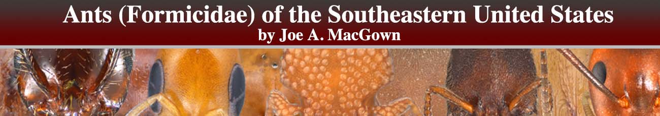
4(3). Mesosomal dorsum lacking promesonotal suture, metanotal suture usually lacking (metanotal suture present in P. californicum), (Fig. 4A); abdominal tergite IV greatly enlarged, strongly arched, in profile view it forms the rearmost part of the abdomen and covers tergites V–VI; apex of abdomen directed anteriorly Fig. 4B) ...Proceratiinae
- Promesonotal suture present; metanotal suture usually present (Fig. 4C); abdominal tergite IV not strongly enlarged, weakly arched, not covering tergites V–VI; apex of abdomen usually directed rearwards or ventrally, not anteriorly (Fig. 4D) ...5
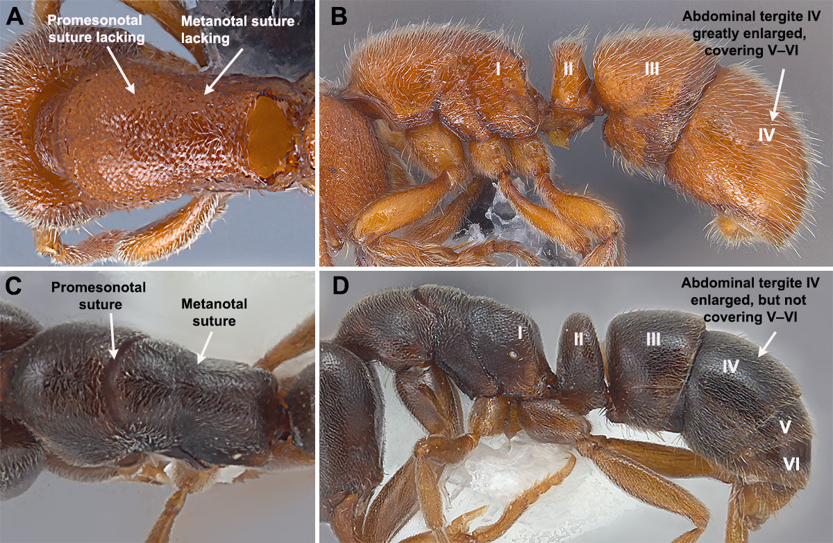 Figure 4. (A) Dorsal view of mesosoma of Proceratium silaceum (Proceratiinae) showing lack of promesonotal and metanotal sutures (B) thorax and abdomen of P. silaceum showing the greatly enlarged abdominal tergite IV (gastral tergite II); (C) dorsal view of mesosoma of H. opacior with arrows pointed toward promesonotal and metanotal sutures, and (D) thorax and abdomen of H. opacior with arrow pointing to abdominal tergite IV (gastral tergite II), which is enlarged but not forming the rearmost part of gaster.
5(4). Head and body covered with deep transverse striae (grooves) (Fig.5A); mandibles elongate and edentate (Fig. 5B) ...Ectatomminae
- Head and body lacking or with limited striae (Fig. 5C); mandibles variable, usually with teeth, at least apically (Fig. 5D) (Leptogenys lacks teeth on mandibles, but differs from Ectatomminae in that the clypeus is elongate and triangular and the head and body does not have deep grooves) ...Ponerinae

Figure 5. (A) Full face view of Poneracantha triangularis (Ectatomminae) showing longitudinal striae and edentate mandibles, (B) lateral view of P. triangularis, note the entire body has deep transverse striae, (C) full face view of Brachyponera chinensis showing the lack of sculpture on the head and the dentate mandibles, and (D) lateral view of B. chinensis showing the general lack of sculpture, especially deep striae, on the body.
6(2). The apex of the gaster with a semicircular to circular acidopore formed from the hypopygium, this structure often projecting as a nozzle and usually with a fringe of setae (Fig. 6A) …Formicinae
- Apex of gaster with hypopygium lacking an acidipore (Fig. 6B) …Dolichoderinae
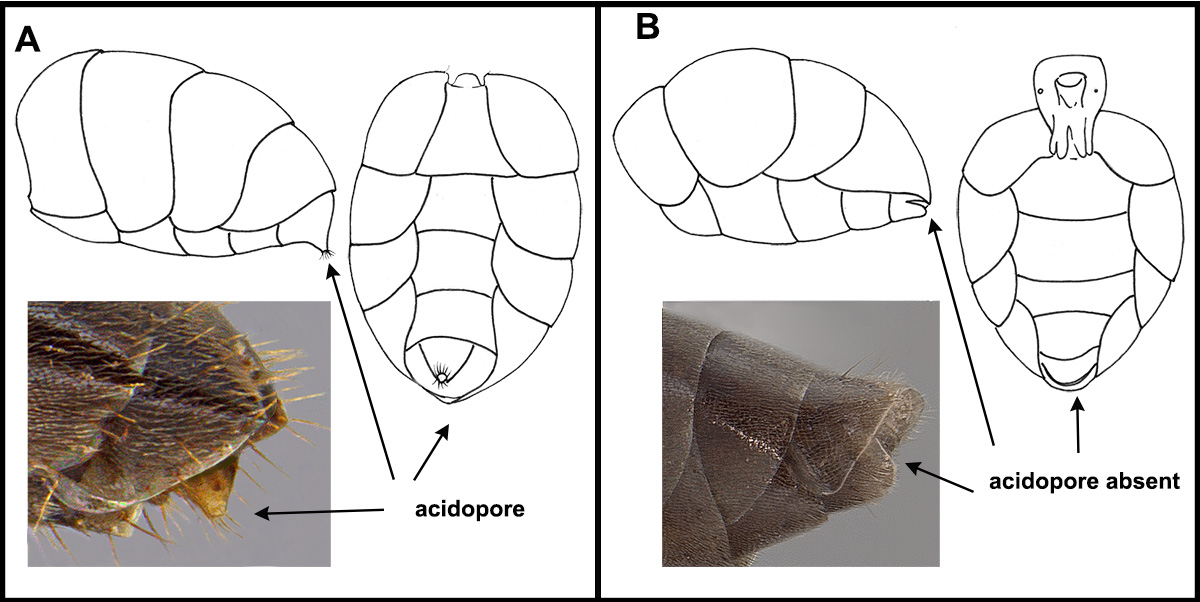
Figure 6. (A) Gastral views of Formicinae showing acidopore and (B) gastral views of Dolichoderinae showing the lack of acidopore.
7(1). Frontal carinae well separated, often covering the bases of the antennae; antennal sockets well separated, set back from anterior border of head; clypeus well developed, extending between frontal carinae (Fig. 6A) ........................................................................Myrmicinae
- Frontal carinae close together, not covering the base of the antennae; antennal sockets close together, near anterior border of head; clypeus reduced (Fig. 6B) ………..............................................................................................................................................................……….8
Figure 7. (A) Full face view of Solenopsis invicta X richteri (Myrmicinae) showing frontal carina being well separated, antennal sockets well separated and set back from anterior margin of head, and clypeus well developed; and (B) full face view of Pseudomyrmex ejectus (Pseudomyrmicinae) showing frontal carinae being close together, antennal sockets well separated and near anterior margin of head, and clypeus reduced.
8(7). Eyes large, reniform, or subelliptical, about half as long as the side of the head; ocelli usually present; antennal scape relatively short, but not unusually thickened and widened, ≈ one-fourth as wide as long, width of scape apically about the same width as pedicel apically (Fig. 7A) …Pseudomyrmecinae
- Eyes with a single facet or absent; ocelli usually absent; antennal scape short and noticeably widened and thickened, one-third to one half (or more) as wide as long, width of scape apically twice as wide or more than width as pedicel apically (Fig. 7B) …Dorylinae
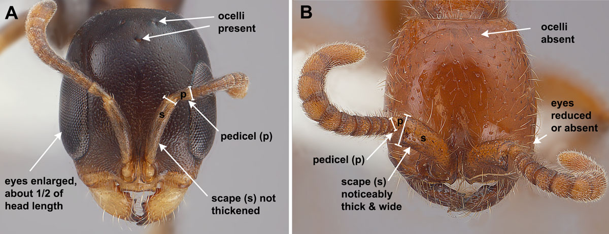 Figure 8. (A) Full face view of Pseudomyrmex ejectus showing enlarged eyes, ocelli, and antennal scapes (not unusually thickened), width of scape apically about the same width as pedicel apically, and (B) Neivamyrmex carolinensis (Dorylinae) showing the lack of eyes and ocelli, and noticeably thickened antennal scapes, width of scape apically twice as wide or more than width as pedicel apically.